Fundamentals of H&E Staining

The aim of this downloadable training resource is to help explain the fundamentals of H&E, or Routine Staining as it’s often termed.
Routine H&E staining plays a critical role in tissue-based diagnosis or research. By coloring otherwise transparent tissue sections, this stain allows highly trained pathologists and researchers to view, under a microscope, tissue morphology. This information is often sufficient to allow a disease diagnosis.
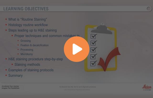
Learn more about:
- What is “Routine Staining”
- Histology routine workflow
- Steps leading up to H&E staining
- Proper techniques and common mistakes in
- Processing
- Microtomy
- Proper techniques and common mistakes in
- Science of H&E staining
- Staining procedure methods
- Staining methods
- Staining dyes and ancillaries
- Examples of staining protocols
- Standard
- Using xylene substitutes
- For frozen sectioning
About the presenter

Andrew Lisowski has almost 30 years of experience in histology and histotechnology. He attended veterinary school and earned his master’s degree in molecular biology. Andrew worked in histology, IHC and ISH labs, cell culture lab, performed in-vitro and in-vivo toxicology assays and was a member of a necropsy team. He worked for pharmaceutical companies, medical school and founded his own molecular and histology firms.
Related Content
Leica Biosystems Knowledge Pathway content is subject to the Leica Biosystems website terms of use, available at: Legal Notice. The content, including webinars, training presentations and related materials is intended to provide general information regarding particular subjects of interest to health care professionals and is not intended to be, and should not be construed as, medical, regulatory or legal advice. The views and opinions expressed in any third-party content reflect the personal views and opinions of the speaker(s)/author(s) and do not necessarily represent or reflect the views or opinions of Leica Biosystems, its employees or agents. Any links contained in the content which provides access to third party resources or content is provided for convenience only.
For the use of any product, the applicable product documentation, including information guides, inserts and operation manuals should be consulted.
Copyright © 2024 Leica Biosystems division of Leica Microsystems, Inc. and its Leica Biosystems affiliates. All rights reserved. LEICA and the Leica Logo are registered trademarks of Leica Microsystems IR GmbH.


