7 result(s) for 'Ki67'
1 - 7 of 7 results for 'Ki67'
Sort by
Show
Antigen Background
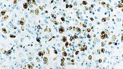
The Ki67 antigen is a nuclear protein which is expressed in all active parts of the cell cycle (G1, S, G2 and mitosis) but is absent in resting cells (G0). In contrast to many other cell cycle-associated proteins, the Ki67 antigen is consistently absent in quiescent cells and is not detectable during DNA repair processes. Thus, the presence of Ki67 antigen is strictly associated with the cell cycle and confined to the nucleus, ...
Phenotyping Tumor Microenvironment Using DNA Barcoded Multiplex Technology Chifei Sun GSK IVIVTBIBCellular Biomarkers Collegeville, PA
I have no conflict of interests with Leica Biosystems, Ultivue and Indica Labs. – All the materials used in this presentation are for educational purpose only. – The human biological samples were sourced ethically, and their research use was in accord with the terms of the informed consents under an IRB/EC approved protocol. Statement 9/25/2020 2
Understand the complexity of tumor microenvironment and its importance in drug development – Introduce the Ultivue DNA barcoding and exchange technology for multiplex immunofluorescence – Highlight the processes of image fusion, spatial analysis and colocalization of markers on individual cells Learning...
Slide 1
Hello, hello; thank you so much for that introduction. Let's get started. Oh, I'm sorry, I just need to hit this next button here. So my name is Mark Lawson.
Thank you for joining us in this webinar. I'm here to present BOND RX tips, tricks, and optimization. It's going to be a user guide from the BOND RX and chromogenic multiplexing in the research application.
Slide 2
So, my name is Mark Lawson. I'm an application specialist on the Life Sciences team at Leica Biosystems I provide technical support for the Life Sciences portfolio, including but not limited to the BOND RX, the BOND RXm, and a wide array of reagents. So I've worked in the histology field for about 15 years and in both clinical and research spaces. I started off as a histotechnologist and worked my way up...
It’s Time for TIME Alexander “Sandy” Borowsky Professor of Pathology and Laboratory Medicine Center for Immunology and Infectious Diseases
Disclosures Bristol-Meyers Squibb/Celgene: Funded through Investigator Sponsored Research (ISR) program. Discovery proposal. Leica/Aperio: Funded to run the “Genesis” study for primary diagnosis from whole slide images. Honorarium for this presentation. Histolix: Slide-free histology (Levenson and Fereidouni). Pfizer: Pfizer Protocol A9001502, Treatment Resistance Following Therapies (TRANSLATE) site PI. Agenda: In contracting to join the “FLEX” registry. None of these has actually paid me, only funding for the research projects.
Objectives • Introduction into the complexity of the TIME and the development of new cancer treatments though di...
Biomarkers
AE1/AE3 (1)
AMACR (2)
BCL6 (1)
CD163 (2)
CD20 (2)
CD3 (9)
CD4 (5)
CD68 (5)
CD8 (10)
CDX2 (2)
CK20 (5)
Ck5 (4)
...
Different types of cancers frequently metastase to bone tissue. Treatment planning decisions are often based upon histology and special staining of these distant sites of disease. These decisions may rely on the outcome of immunohistochemistry, in situ...
The content of this webinar will encompass detailed technical descriptions of novel and state of the art methods to decalcify tissue samples that will be embedded in paraffin or for frozen sectioning for molecular and nucleic acid analysis for basic research...