Utility of Adequate Core Biopsy Samples from Ultrasound Biopsies Needed for Today’s Breast Pathology
Abstract
Background: There is a paradigm shift in breast biopsy philosophy. In the past radiologists and clinicians used to collect as little tissue as possible for pathologists to render a diagnosis on conventional histologic H&E sections. Precision medicine has changed this philosophy in such a way that more optimal core biopsy specimens are now required to provide more data for personalizing the therapy.
Methods: A case is presented in this study to illustrate the importance of adequate ultrasound-guided breast biopsy samples. Digital mammography of left breast in a postmenopausal woman revealed a spiculated lesion, which was confirmed with ultrasonography as a 0.4 cm irregular mass. A core biopsy was performed by using an ultrasound-guided 13 gauge tetherless vacuum-assisted Mammotome Elite biopsy system. Eleven cores were submitted for paraffin embedding in four cassettes. Following microscopic examination of histologic slides, additional diagnostic and prognostic ancillary studies were performed.
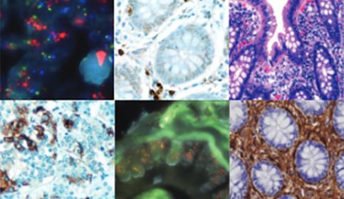
Results: Microscopic examination revealed a moderately-differentiated invasive ductal carcinoma with a maximum histologically contiguous linear extent of 0.4 cm, and low to intermediate grade DCIS (DCIS grade 1-2) without calcifications. Radiological and core biopsy carcinoma dimensions of the lesion were concordant. The core biopsy sample displayed the entire invasive carcinoma which consisted of well-fixed, abundant invasive carcinoma tissue for all ancillary studies.
Conclusions: Ultrasound-guided core biopsies obtained using tetherless Mammotome Elite system provide high quality, unfragmented, adequate samples when a breast mass is targeted. Routine ancillary studies, prognostic panels, and advanced comprehensive genomic testing are performed with ease on these adequate samples.
Given the heterogeneous nature of breast carcinomas, more sufficient sampling of breast masses not only enables the pathologist to characterize the tumor adequately, but also provides better-represented, adequate tumor tissue for comprehensive genomic testing for personalized medicine. Mammotome Elite samples could easily provide optimum tumor surface area (25 mm2) and tumor volume (2mm3) for such genomic testing for rendering a confident diagnosis.
If you have viewed this educational webinar, training or tutorial on Knowledge Pathway and would like to apply for continuing education credits with your certifying organization, please download the form to assist you in adding self-reported educational credits to your transcript.
Apply for self-reported educational creditsIntroduction
Histopathologic examination of adequate, minimallyinvasive breast core biopsy samples corresponding to an abnormal radiological finding is the standard of care prior to any therapeutic intervention or mammographic surveillance decision. Excision of breast (breast conserving surgery, mastectomy) without prior minimally invasive core biopsy diagnosis is almost an obsolete clinical practice.
It is imperative to have a quantitatively and qualitatively adequate sample in order for a pathologist to formulate an ironclad diagnosis. Majority of patients with imaging abnormality who have undergone minimally-invasive breast core biopsy with benign diagnosis are spared further excision and excision-related complications. Many of these patients are managed with radiological surveillance only.
Quality of the individual tissue sample should be optimal to provide a confident radiological-pathological correlation. Undersampling of clinically significant lesions (invasive / in situ carcinoma, atypical ductal hyperplasia, complex radial scars, papillary neoplasms with or without atypia) and fragmented inadequate samples could easily lead to a false negative “benign breast tissue” histologic diagnosis. According to a recent article published in the Journal of Clinical Oncology, insufficient cancer tissue for biomarker testing occurred across 4 out of 5 cancer types reported on. The majority of tissue samples in which insufficient tissue was present were acquired through core biopsies (67% of all cases) or FNAs (22% of all cases).1
Quantity of the sample should be optimal to perform ancillary diagnostic studies (P63, calponin, CK903, E-cadherin, CK5/6, CK 7, MNF 116, S100, collagen type IV, reticulin stains etc.) or prognostic predictive markers (ER, PR, AR, Ki67,