10 result(s) for 'CD68'
1 - 10 of 10 results for 'CD68'
Sort by
Show
Antigen Background
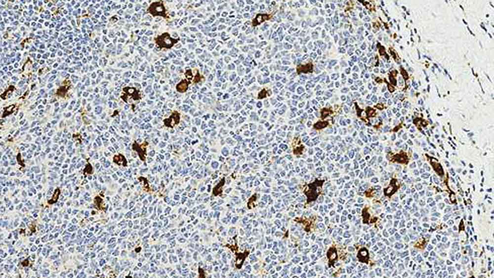
CD68は、主としてマクロファージの細胞質顆粒に(より低レベルで細胞膜にも)結合すると報告されている細胞内糖タンパク(110kD)です。CD68は、マクロファージの同定に最も高頻度に利用されている免疫組織化学マーカーですが、単球、好中球、好塩基球および大型リンパ球にもみられます。CD68の機能は明らかにされていませんが、酸性加水分解酵素からライソゾーム膜を保護している可能性があります。細胞表面のCD68に機能的な意義があるのか、またはライソゾームからの漏出によるものなのかは明らかにされていません。CD68の発現は、刺激されたT細胞、NK細胞、肝臓や尿細管などの非造血系組織でも証明されています。
Disclaimer
CD68 is recommended for the detection of specific antigens of interest in normal and neoplastic tissues, as an adjunct ...
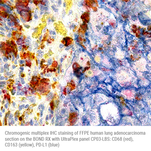
DESCRIPTION: Chromogenic multiplex IHC kit for BOND RX - human CD68, CD163, PD-L1
SIZE: 10 slides
PRODUCT OVERVIEW: Kit for chromogenic multiplex IHC staining of 10 slides plus a single negative control slide on the BOND RX with V7.0 software.
1° Antibody
Clone
1° Tag
2° Antibody
Chromogen
CD8
C68/684
UT021
Anti-UT021-AP
Leica Refine Red
CD163
EPR19518
UT015
Anti-UT015-HRP
Yellow
PD-L1
73-10
UT014
Anti-UT014-HRP
Leica Blue
Leica Refin...
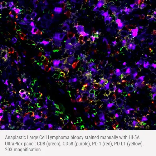
DESCRIPTION: Kit for fluorescent multiplex IHC staining of human CD8, CD68, PD-1, PD-L1, detection with 490, 550, 650, 750 nm fluors
SIZE: 5 slides
PRODUCT OVERVIEW: All components required to stain 5 multiplex slides plus a single negative control slide with simple two-step protocol
1° Antibody
Clone
1° Tag
2° Antibody
2° Fluor
CD8
EPR10640(2)
UT015
Anti-UT015
CL490
CD68
C68/684
UT019
Anti-UT019
CL750
PD-1
EPR4877
UT016
Anti-UT016
CL650...
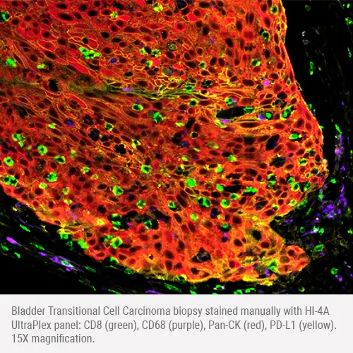
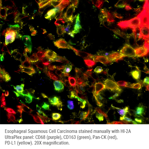
DESCRIPTION: Kit for fluorescent multiplex IHC staining of human CD8, CD68, Pan-CK, PD-L1, detection with 490, 550, 650, 750 nm fluors
SIZE: 5 slides
PRODUCT OVERVIEW: All components required to stain 5 multiplex slides plus a single negative control slide with simple two-step protocol.
1° Antibody
Clone
1° Tag
2° Antibody
2° Fluor
CD8
EPR10640(2)
UT015
Anti-UT015
CL490
CD68
C68/684
UT019
Anti-UT019
CL750
Pan-CK
AE1/AE3
UT016
Anti-UT016
...
DESCRIPTION: Kit for fluorescent multiplex IHC staining of human CD68, CD163, Pan-CK, PD-L1, detection with 490, 550, 650, 750 nm fluors
SIZE: 5 slides
PRODUCT OVERVIEW: All components required to stain 5 multiplex slides plus a single negative control slide with simple two-step protocol.
1° Antibody
Clone
1° Tag
2° Antibody
2° Fluor
CD6
C68/684
UT019
Anti-UT019
CL750
CD163
EPR19518
UT015
Anti-UT015
CL490
Pan-CK
AE1/AE3
UT016
Anti-UT016
...
Phenotyping Tumor Microenvironment Using DNA Barcoded Multiplex Technology Chifei Sun GSK IVIVTBIBCellular Biomarkers Collegeville, PA
I have no conflict of interests with Leica Biosystems, Ultivue and Indica Labs. – All the materials used in this presentation are for educational purpose only. – The human biological samples were sourced ethically, and their research use was in accord with the terms of the informed consents under an IRB/EC approved protocol. Statement 9/25/2020 2
Understand the complexity of tumor microenvironment and its importance in drug development – Introduce the Ultivue DNA barcoding and exchange technology for multiplex immunofluorescence – Highlight the processes of image fusion, spatial analysis and colocalization of markers on individual cells Learning...
Slide 1
Hello, hello; thank you so much for that introduction. Let's get started. Oh, I'm sorry, I just need to hit this next button here. So my name is Mark Lawson.
Thank you for joining us in this webinar. I'm here to present BOND RX tips, tricks, and optimization. It's going to be a user guide from the BOND RX and chromogenic multiplexing in the research application.
Slide 2
So, my name is Mark Lawson. I'm an application specialist on the Life Sciences team at Leica Biosystems I provide technical support for the Life Sciences portfolio, including but not limited to the BOND RX, the BOND RXm, and a wide array of reagents. So I've worked in the histology field for about 15 years and in both clinical and research spaces. I started off as a histotechnologist and worked my way up...
Biomarkers
AE1/AE3 (1)
AMACR (2)
BCL6 (1)
CD163 (2)
CD20 (2)
CD3 (9)
CD4 (5)
CD68 (5)
CD8 (10)
CDX2 (2)
CK20 (5)
Ck5 (4)
...
It’s Time for TIME Alexander “Sandy” Borowsky Professor of Pathology and Laboratory Medicine Center for Immunology and Infectious Diseases
Disclosures Bristol-Meyers Squibb/Celgene: Funded through Investigator Sponsored Research (ISR) program. Discovery proposal. Leica/Aperio: Funded to run the “Genesis” study for primary diagnosis from whole slide images. Honorarium for this presentation. Histolix: Slide-free histology (Levenson and Fereidouni). Pfizer: Pfizer Protocol A9001502, Treatment Resistance Following Therapies (TRANSLATE) site PI. Agenda: In contracting to join the “FLEX” registry. None of these has actually paid me, only funding for the research projects.
Objectives • Introduction into the complexity of the TIME and the development of new cancer treatments though di...
The content of this webinar will encompass detailed technical descriptions of novel and state of the art methods to decalcify tissue samples that will be embedded in paraffin or for frozen sectioning for molecular and nucleic acid analysis for basic research...