13 result(s) for 'CD3'
1 - 10 of 13 results for 'CD3'
Sort by
Show
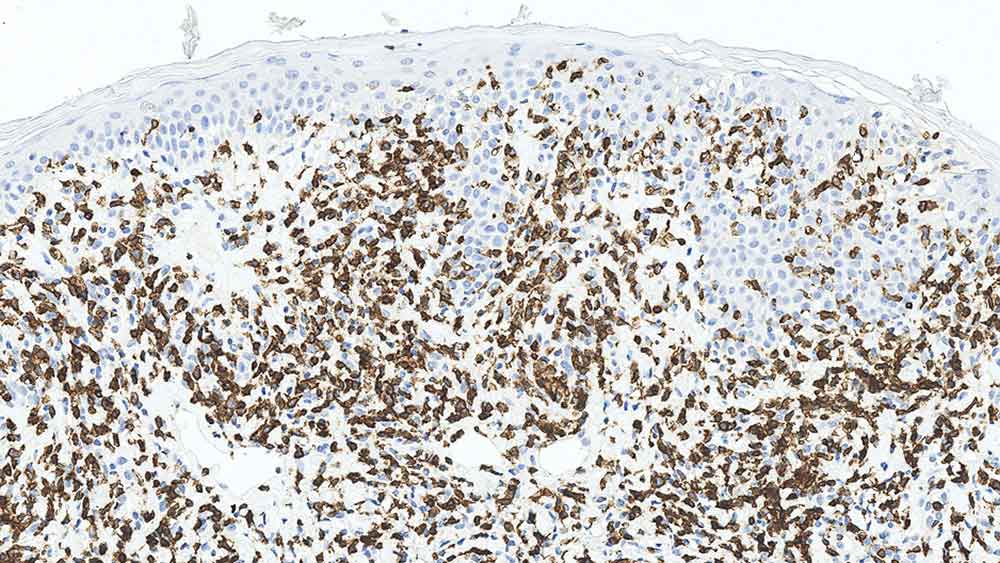
Antigen Background
CD3 は、分子量16-28kDの5 種類のポリペプチド鎖からなります。CD3 は、初期胸腺細胞で最初に検出され、その出現はT細胞系への分化系統決定を示す最も早い段階でのサインの1つであると考えられています。胸腺、骨髄、末梢リンパ組織および血液におけるT細胞を認識する汎T細胞マーカーです。
Product Specific Information
Clone LN10 抗体は、ヒトCD3分子の非グリコシル化Epsilon鎖に特異的です。
Disclaimer
CD3 is recommended for the detection of specific antigens of interest in normal and neoplastic tissues, as an adjunct to conventional histopathology using non-immunologic histochemical stains....
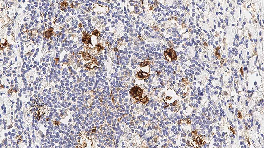
Antigen Background
CD30は、分子量120kDの単鎖糖タンパクです。CD30は、サイトカインのリガンドであるCD30Lの受容体として作用することが知られており、細胞増殖の調節と形質転換においても役割を果たしている可能性があります。CD30は、ReedSternberg細胞、ホジキン細胞および大部分の未分化大細胞型リンパ腫の細胞表面に発現することが報告されています。また、CD30は、非ホジキンリンパ腫およびウイルスによる形質転換細胞(EBV)により形質転換されたB細胞などに発現しています。
Product Specific Information
Clone JCM182 抗体はIFUにて推奨されている溶液(pH6.0)以外の抗原賦活液を使用すると、背景染色が増強することがあります。
Disclaimer
CD30 is recommended for the detection of specific antigens of interest in normal an...
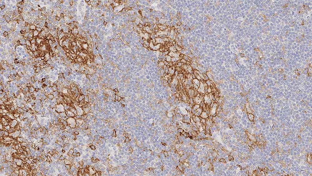
Antigen Background
CD33は、骨髄単球性前駆体細胞でCD34の後に発現することが報告されています。骨髄系細胞および単球系細胞に発現しますが、顆粒球には存在しないことが報告されています。CD33の発現は、単球、前骨髄球、骨髄芽球、一部の未分化型急性白血病、急性リンパ芽球性白血病に限定されると報告されています。CD34
Disclaimer
CD33 is recommended for the detection of specific antigens of interest in normal and neoplastic tissues, as an adjunct to conventional histopathology using non-immunologic histochemical stains.
NCL-L-CD38-290,CD38,SPC32,1mL
---JC70A---PA0414.jpg)
NCL-L-CD31-607,CD31,JC70A,1mL
---QBEnd-10---PA0212.jpg)
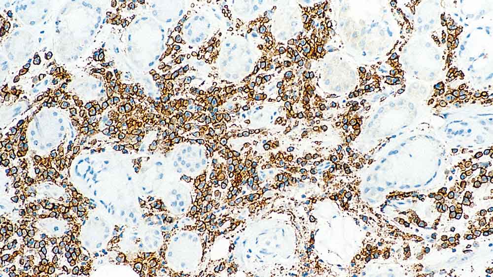
Antigen Background
CD34は、分子量110kDの単鎖膜貫通型糖タンパクです。CD34は、ヒトリンパ系および骨髄系前駆細胞に選択的に発現しています。また、血管内皮にも発現しています。
Product Specific Information
Enzyme digestion of paraffin sections is recommended with clone QBEnd/10 in perference to heat induced epitope retrieval as it produces stronger staining and reduces background elastin staining
Disclaimer
CD34 (Endothelial Cell Marker) is recommended for the detection of specific antigens of interest in normal and neop...
PA0414,CD31,JC70A,7mL
【中止1610】CD31 (PECAM-1)
It’s Time for TIME Alexander “Sandy” Borowsky Professor of Pathology and Laboratory Medicine Center for Immunology and Infectious Diseases
Disclosures Bristol-Meyers Squibb/Celgene: Funded through Investigator Sponsored Research (ISR) program. Discovery proposal. Leica/Aperio: Funded to run the “Genesis” study for primary diagnosis from whole slide images. Honorarium for this presentation. Histolix: Slide-free histology (Levenson and Fereidouni). Pfizer: Pfizer Protocol A9001502, Treatment Resistance Following Therapies (TRANSLATE) site PI. Agenda: In contracting to join the “FLEX” registry. None of these has actually paid me, only funding for the research projects.
Objectives • Introduction into the complexity of the TIME and the development of new cancer treatments though di...
Phenotyping Tumor Microenvironment Using DNA Barcoded Multiplex Technology Chifei Sun GSK IVIVTBIBCellular Biomarkers Collegeville, PA
I have no conflict of interests with Leica Biosystems, Ultivue and Indica Labs. – All the materials used in this presentation are for educational purpose only. – The human biological samples were sourced ethically, and their research use was in accord with the terms of the informed consents under an IRB/EC approved protocol. Statement 9/25/2020 2
Understand the complexity of tumor microenvironment and its importance in drug development – Introduce the Ultivue DNA barcoding and exchange technology for multiplex immunofluorescence – Highlight the processes of image fusion, spatial analysis and colocalization of markers on individual cells Learning...