
When to Use a Vibrating Microtome for Tissue Sectioning
Users of microtomes often ask if they can use just one microtome across all functions in the laboratory. This article addresses the advantages and limitations of Vibrating Blade microtomes.
Other microtome types require hardening the tissue by freezing or paraffin embedding. This is because a blade, no matter how sharp, pushed forward into a block of soft tissue, will compress the tissue in front of it rather than cut. When compressed enough to push back, the tissue will suddenly cut, but not straight through, the blade bevel and uneven compression will create deflections. However, soft tissue can be cut by rapidly moving the blade back and forth, thus creating a sawing effect as the blade slowly advances forward. Why cut soft?
Advantages of a Vibrating Microtome:
- Does not fracture cell membranes (freezing frequently does); cytosol remains in cells.
- Stains are sharper, with no cytosol in the extracellular tissue. Better for HRP, etc.
- Can keep cells living. Tissue may be oxygenated under media while cutting.
- Quick setup, glue tissue to the platform. No encapsulation is needed.
Disadvantages of a Vibrating Microtome:
- Much slower to operate than a cryostat or rotating or sliding microtome.
- Sections must be thick, 50 microns and up.
- Block from which sections are taken must not be too thick.
Issues encountered in Vibrating Microtomy:
Blade Angle:
The blade can be set to any angle. The correct blade angle depends only on the properties of the blade. However, the consequences of incorrect blade angle are different depending on the properties of the material being cut as illustrated below.
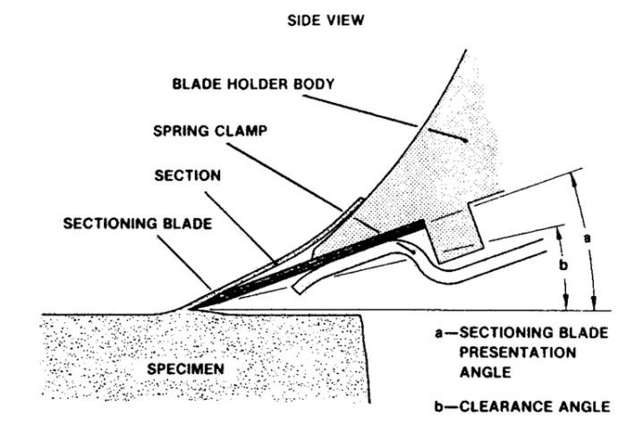
This image from an old manual actually shows an incorrect blade angle and clearance angle. See the article Knife Angle in Microtomy. The "Blade Angle" is the angle between the blade body and the direction of motion. Note that there is a bevel on the front edge of the blade. This is necessary for blade manufacturing. The clearance angle is the angle between the lower bevel and the direction of motion. The blade angle forces the tissue to bend upward at the blade front. This compresses cells on one side of the section at the bend and stretches them on the other. This distortion should always be minimized. However, if the blade angle is zero, the clearance angle will be negative because the bevel is still there. The back of the lower bevel will push down and compress the block as it passes over and slide along the tissue. This is highly undesirable for any application. To avoid this risk, the clearance angle, the angle between the bevel on the blade and the path of advance, must be above zero. At zero, there would still be some friction. An angle of about 1° would be perfect. There is no case where more than that is advantageous. Note in the imagethat it is about 10-15 degrees, and thus bending the tissue more than is necessary. The correct blade angle is thus dependent on, and slightly greater than, the bevel angle of the blade being used, and no other property.
Amplitude of Oscillation
For mechanical reasons having to do with the natural frequency of oscillation, it is not mechanically advisable to speed up the back and forth motion over the same range. However, by increasing the amplitude of oscillation, with each oscillation still at the same frequency, one can have the blade moving faster against the tissue part of the time. As long as the greater amplitude is not tearing the tissue at the extremes or causing vertical vibration of the blade, more is better, especially for tissue that wants to compress.
Speed of Forward Advance
If the blade is slipping out of the tissue block or compressing the tissue, try a slower advance. For the most critical applications, such as calcium dual fluorescent imaging in live tissue sections, the slower, the better, some use 10 minutes per section, as long as the tissue is still alive when you have the section. The trade-off is the speed of getting the job done .versus section quality.
Thick or Thin Sections
If the instrument is cutting alternate sections of different thickness, there are a few possible causes. Clearance angle negative is a common one; the blade angle is too low. Another is something loose; the pedestal, the tissue on it, or the blade is not tightly secured. Try to grasp and wobble things one at time, preferably without cutting yourself. Another possible cause is that the tissue has a high height for the base of support, and is flexing at the top, so the tissue is moving. Cut the block smaller if feasible or find a way to support the tissue better.
Related Content


El contenido de Leica Biosystems Knowledge Pathway está sujeto a las condiciones de uso del sitio web de Leica Biosystems, disponibles en: Aviso legal.. El contenido, incluidos los webinars o seminarios web, los recursos de formación y los materiales relacionados, está destinado a proporcionar información general sobre temas concretos de interés para los profesionales de la salud y no está destinado a ser, ni debe interpretarse como asesoramiento médico, normativo o jurídico. Los puntos de vista y opiniones expresados en cualquier contenido de terceros reflejan los puntos de vista y opiniones personales de los ponentes/autores y no representan ni reflejan necesariamente los puntos de vista ni opiniones de Leica Biosystems, sus empleados o sus agentes. Cualquier enlace incluido en el contenido que proporcione acceso a recursos o contenido de terceros se proporciona únicamente por comodidad.
Para el uso de cualquier producto, debe consultarse la documentación correspondiente del producto, incluidas las guías de información, los prospectos y los manuales de funcionamiento.
Copyright © 2024 Leica Biosystems division of Leica Microsystems, Inc. and its Leica Biosystems affiliates. All rights reserved. LEICA and the Leica Logo are registered trademarks of Leica Microsystems IR GmbH.