23 result(s) for 'Colon'
1 - 10 of 23 results for 'Colon'
Sort by
Show
---EPMU1---PA0210.jpg)
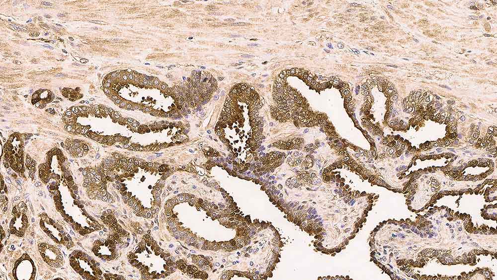
Antigen Background
Alpha-methylacyl-CoA racemase (AMACR), also known as p504s, is a mitochondrial and peroxisomal enzyme that is involved in bile acid biosynthesis and beta-oxidation of branched-chain fatty acids. AMACR is essential in lipid metabolism, and is expressed in normal liver (hepatocytes), kidney (tubular epithelial cells) and gall bladder (epithelial cells). Expression has also been found in lung (bronchial epithelial cells) and colon...
---COL-1---PA0848.jpg)
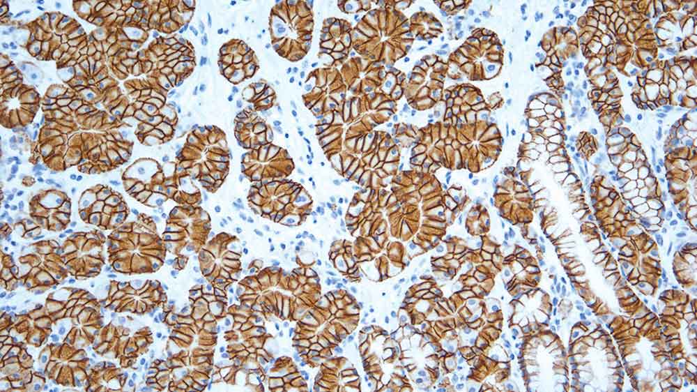
Antigen Background
Carcinoembryonic antigen (CEA) is a heterogeneous cell surface glycoprotein produced by cells of fetal colon. Low levels are also found on normal mucosal epithelia of the adult colon and a variety of other normal tissues. CEA is encoded by the CEA gene, which is located on chromosome 19. It is a member of the CEA gene family, which in turn is a subfamily of the immunoglobulin superfamily. Cell adhesion properties are now well r...
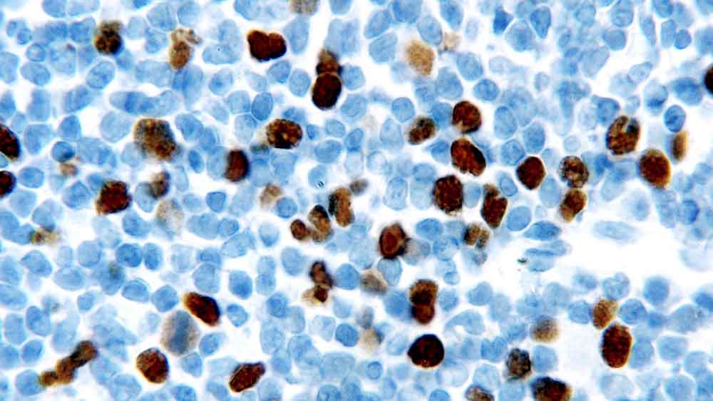
Antigen Background
Geminin is a protein of 209 amino acids thought to be involved in the control of DNA replication via the interaction with Cdt1. Geminin is not found in the G1 phase of the cell cycle, but is first expressed in the G1 to S transition phase, with expression levels rising through the rest of the cell cycle and levels reaching a maximum during mitosis. It has been proposed that geminin may be a tumor suppressor protein. Geminin is ...
Antigen Background
Carbonic anhydrase (CA) is an enzyme that assists rapid interconversion of carbon dioxide and water into carbonic acid, protons, and bicarbonate ions. Originally named MN/G250, carbonic anhydrase IX (CAIX) is a cell surface transmembrane protein, which is predominantly found in the gastrointestinal tract and gallbladder. The glandular regions of normal colon are reported to be negative, but in the case of adenocarcinoma, the gl...
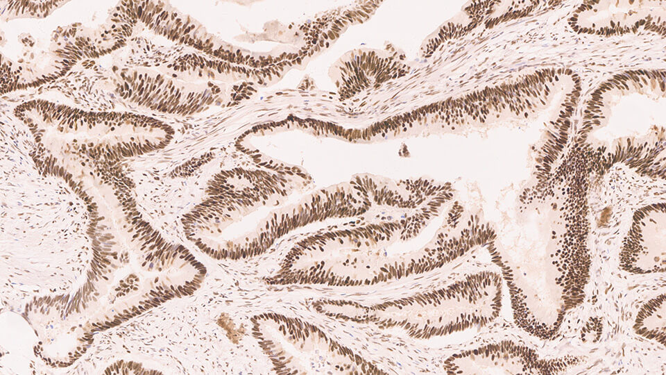
Antigen Background
MSH6 is a 160 kD protein which is involved in DNA mismatch repair (MMR) and recombination pathways, when heterodimerized with MSH2. Defects in mismatch repair systems can cause mutations and can cause DNA microsatellite sequences to become unstable. Immunohistochemical studies have reported that MSH6 is strongly expressed in the nucleus of cells in normal colonic epithelium, especially in crypts. Expression is also found in lym...
Antigen Background
Galectin-3 is a member of the beta-galactosidase-binding lectin family. It is involved in several biological events including binding to the basement membrane glycoprotein laminin. Cell surface galectin-3 may be involved in homotypical cell adhesion and is downregulated in colon cancer as the disease progresses. This downregulation has also been examined in breast carcinoma with a similar correlation of expression reported. Dow...
Antigen Background
MLH1, a mismatch repair protein involved in maintaining the integrity of genetic information, alongside MSH2, MSH6 and PMS2. During DNA replication, strand misalignment can occur resulting in alterations to microsatellite repeats, often referred to as microsatellite instability (MSI). These defects in DNA repair pathways have been linked to human carcinogenesis. Mutations in the MLH1 gene have been reported to be found in some ...

Antigen Background
Human mismatch repair protein 2 (MSH2) is involved in the initial recognition of mismatched nucleotides during the post replication mismatch repair process. Therefore, the loss of MSH2 function leads to the accumulation of replication errors, which in turn may be responsible for the multiple mutations required for multistage carcinogenesis. MSH2 is reported to be expressed in the nuclei of cells from a variety of tissues includ...
Slide 1
Welcome
Slide 2
Slide 3
Discuss Pre-Analytics and the impact of improper fixation and artifacts on tissue processing
Discuss Fixation and the impact of incomplete fixation on tissue processing
Describe the impact of improper Prosection
Determine satisfactory Processing of samples
Explain why routine Maintenance is a critical success factor to proper tissue processing
Slide 4
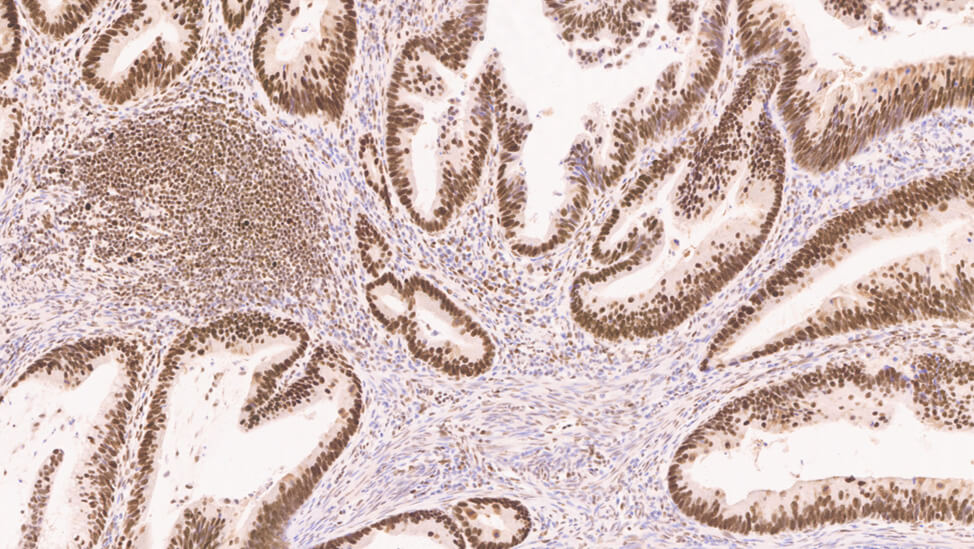
This slide has two examples of what the ideal sections in a perfect world look like under the scope. The skin section on the left is clean looking with the Eosin staining the components of the dermis with different shades. The colon section on the right is crisp with well-defined nuclei and cilia. There is no background staining or muddiness to the stain.
Slide 5
One artifact seen the lab that is n...
One of the most fundamentally critical elements of diagnostic histopathology is first the ability to suspend all cellular activity in tissue and prevent degradation, and secondly to process that specimen in a manner that facilitates subsequent steps such as...