


Aperio GT 450 DX System


Overview
High-Volume Clinical Pathology Scanning Powered by Leica Precision Optics
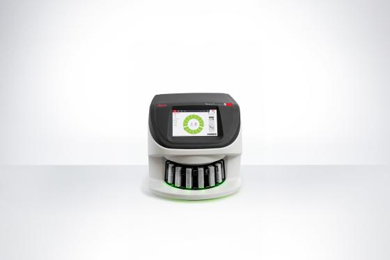
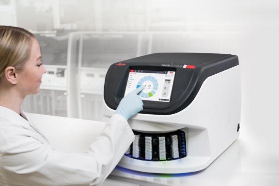
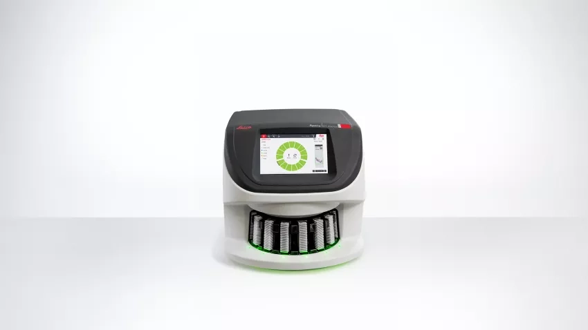
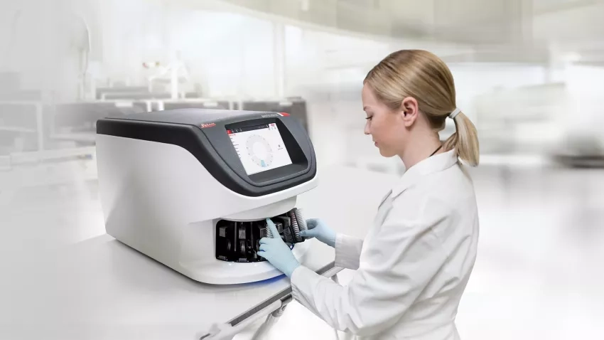
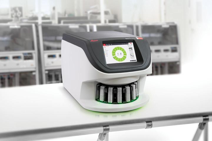
The Aperio GT 450 DX is an automated, high-capacity digital pathology slide scanner engineered to accelerate diagnostic workflows in high-volume laboratories. Combining Leica optics with continuous, no-touch loading, the system delivers high diagnostic clarity at rapid scan speeds.
- * High-Speed Throughput: Scans a standard 15 mm x 15 mm area at 40x magnification in 32 seconds per slide—achieving an operational throughput of 81 slides per hour (per Leica Biosystems validation VP-0450).
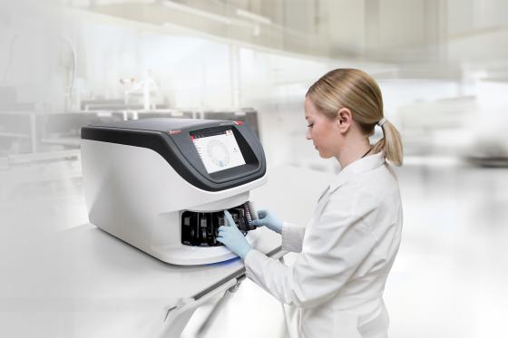
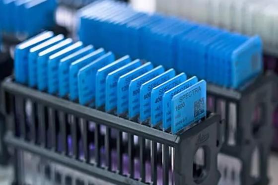
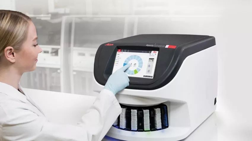
- * Continuous High-Capacity Loading: Holds up to 450 slides across 15 racks. Supports continuous, direct rack transfer from the HistoCore SPECTRA Workstation (Stainer & Coverslipper) without manual handling.
- * Patented Real-Time Focus (RTF): Employs dual-sensor line technology (US Patent No. 9,841,590) to dynamically adjust focal depth on the fly, eliminating focal mapping delays at 40x.
- * Sub-Cellular Resolution: Custom Leica optical objective delivers a wide 1 mm Field of View (FOV) with flat field correction, capturing detailed images at 0.26 µm/pixel resolution.
- * Enterprise Interoperability: Native DICOM-compatible and SVS image output formats with HL7 integration via a centralized Scanner Administrator Manager (SAM) DX server.
- * Automated Operational Precision: Features a 99.5% tissue finder accuracy and a 100% first-time barcode scan success rate (per Leica Biosystems validation VP-0463).
*Optional features to meet your workflow requirements. Some features may not be available in all countries.
For In Vitro Diagnostic Use.
Features
Manual Scan
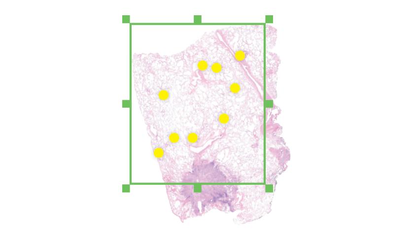
Provides histotechnicians with direct manual overrides to ensure successful high-resolution scanning of difficult or non-standard glass slides:
- * Custom Scan Region Selection: Allows operators to manually define specific regions of interest (ROIs) on faint or small specimen fragments.
- * Manual Focus Calibration: Enables precise manual focus point placement for uneven, weakly stained, or non-standard tissue sections.
- * Workflow Continuity: Prevents challenging slides from stalling automated batch runs, maintaining consistent turnaround times.
Multiplane Imaging & Focal Depth Management
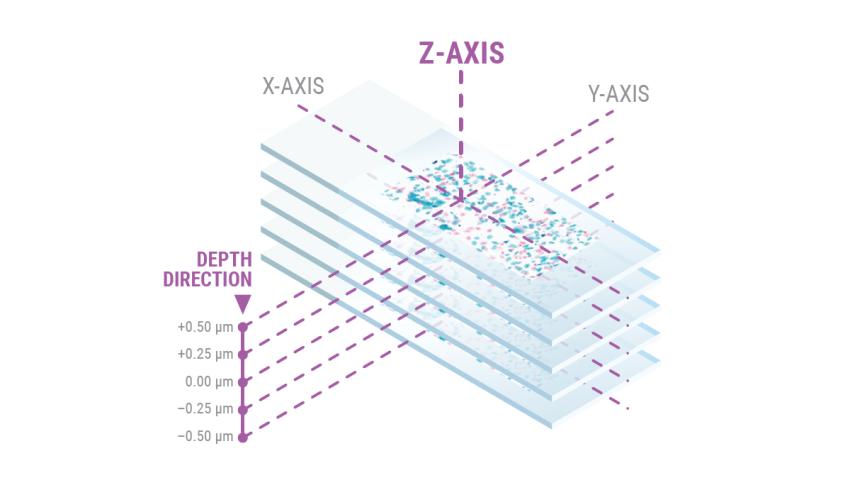
- * Z-Stacking: Scans multiple focal planes across the z-axis and compiles them into a single multiplane image, enhancing diagnostic confidence for thick or multi-layered specimens.
- * Extended Focus: Combines focal planes into a single optimized 2D image layer, maximizing visual sharpness while reducing storage space compared to full Z-stack datasets.
- * Real-Time Focusing (RTF - US Patent No. 9,841,590): Synchronizes an extra focusing sensor with the primary imaging line sensor to adjust objective height dynamically during continuous slide translation.
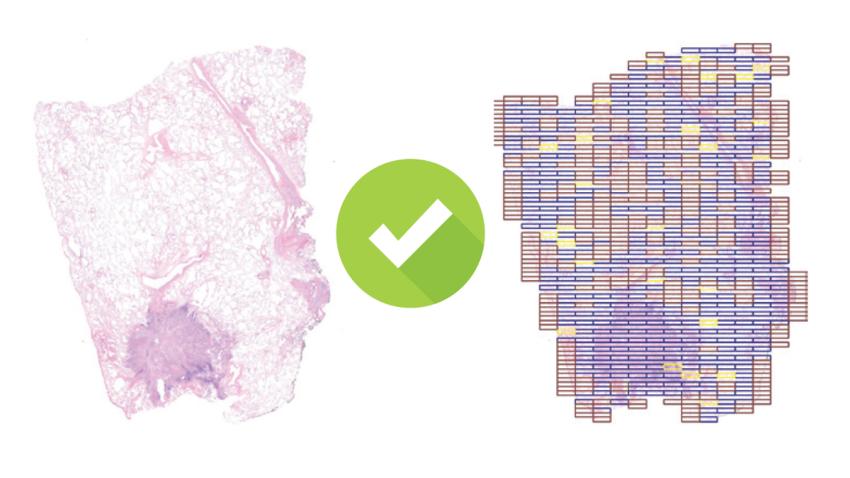
Quality Control & Image Standardization
- * Aperio iQC DX Software (Optional): AI-enabled quality control software that scans whole slide images (WSIs) for visual artifacts, flagging substandard slides on a centralized dashboard before routing cases to pathologists.
- * Default Calibration Point: Pre-set optical calibration parameters reduce optical striping at scan initialization without requiring manual corrective steps.
- * Automated Per-Scan Calibration: Calibrates light intensity and white balance for every slide individually to guarantee image consistency across entire batch runs.
- * Automated Narrow Stripe Functionality: Eliminates image stitch lines and optical artifacts across adjacent scan paths.
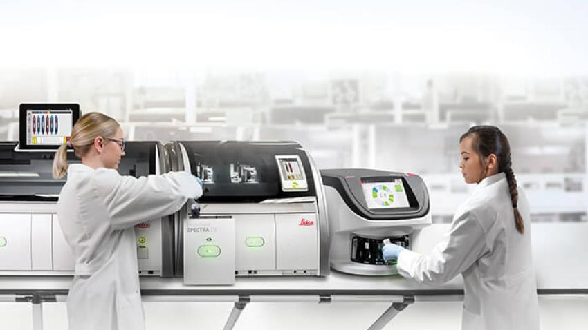
High-Efficiency Lab Operations & Automation
- * Direct Workstation Integration: Accepts 30-slide Leica Universal Racks directly from the HistoCore SPECTRA Coverslipper, eliminating manual slide transfer. (Also supports 20-slide Sakura Prisma racks).
- * Smart Tissue Finder: Validated 99.5% accuracy in distinguishing faint tissue specimens from pen marks, mounting residue, dust, and coverslip edges.
- * 100% Barcode Success Rate: High-accuracy engine supports 1D and 2D barcode symbologies (Data Matrix, QR Code, Code 128, Code 39, PDF417) to prevent patient tracking errors.
- * Priority Case Processing: Allows technicians to flag urgent racks for immediate scanning without interrupting active runs.
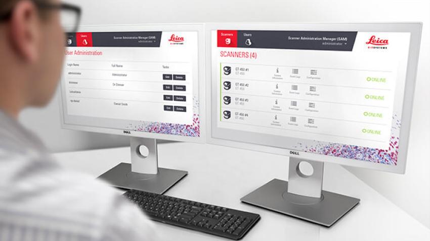
Enterprise IT Architecture & Cybersecurity (SAM DX)
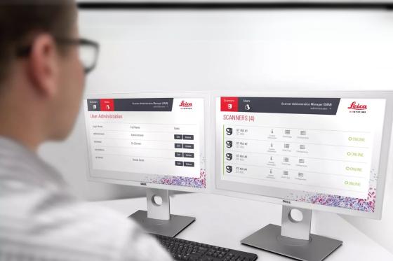
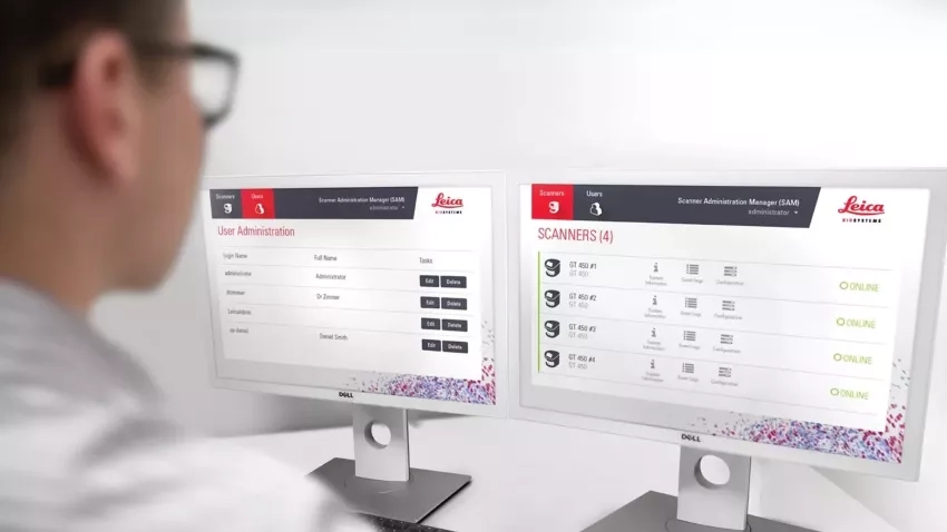
- * Centralized SAM Server: The Scanner Administrator Manager DX enables remote configuration, status monitoring, and management of multiple GT 450 DX units from a single interface.
- * Role-Based Security: Features dedicated scanner logins, user-specific PIN access, custom permission groups, and automatic security timeouts.
- * Network & Data Protocols: Utilizes SSL encryption for server communications, Mirth Connect event logging, HL7 interface engine compatibility, and native DICOM image output to PACS/LIS architectures.
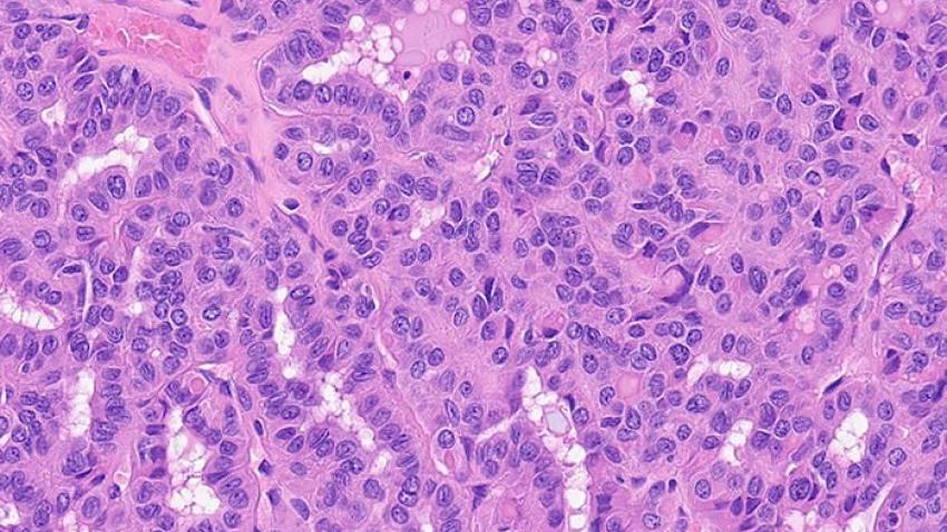
Pathologist-Validated Image Quality
Engineered with custom Leica Microsystems optics (crafted since 1849) to deliver clear, on-screen diagnostic review via Aperio Webviewer DX:
- * Sub-Cellular Clarity: High-contrast resolution optimized for evaluating fine diagnostic detail, including mitotic events, intercellular bridges, chromatin structure, and nucleoli.
- * Real-Time Calibration: Every slide is automatically calibrated during scanning to prevent optical striping and artifacts.
- * Automated Artifact Exclusion: 99.5% accurate tissue finder detects faint tissue while excluding pen marks, dust, and mounting residue to eliminate unnecessary re-scans.
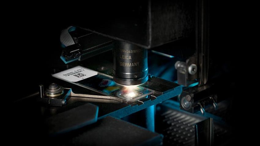
Custom-Made Leica Optics
Custom-engineered objective manufactured by Leica Microsystems (optical craftsmanship since 1849) specifically for high-speed digital pathology scanning:
- * Extra-Wide Field of View: Features a flat-field corrected 1.0 mm Field of View (FOV)—far exceeding standard round objectives—to capture extremely large digital areas for rapid scan speeds.
- * High-Resolution Detail: Delivers 0.26 µm/pixel resolution at 40x magnification, providing on-screen clarity for sub-cellular diagnostic review.
- * Per-Slide Calibration: Automatically calibrates light and white balance during every individual scan to maintain consistent optical performance across large batch runs.
Specifications
Sakura Prisma Stainer and Coverslipper Rack (20-slide capacity)
*VP-0450
(–includes automatic timeouts)
(–password for client software and PIN for scanner)
Slides
Slides are prepared typically using:
• Glass coverslip with mounting media, Eukitt
• Film coverslip with integrated glue
optimised for tissue thickness of 3-5 μm
(including mounting media)
Coverslips
Labels
Barcodes
Downloads
Similar Products

HistoCore SPECTRA Automated Slide Staining Workstation
Knowledge Hub
From tissue processing to Digital Pathology, our solutions empower labs with efficiency, precision, and reliability.
Top Considerations When Buying a Digital Pathology Scanner
Are you thinking about purchasing a digital pathology scanner? We offer some tips for things to consider during the buying process.
Four Tips for Digital Pathology Slide Scanning
Do you want to get better digital images from your glass slides? Here are some tips to help you get the best out of your digital pathology line scanner:
Introducing the Aperio GT 450 DX
81 slides per hour @ 40x (15mm x 15mm area).
32 seconds at 40x for 15mm x 15mm area
No, Aperio GT 450 DX has a scalable IT architecture solution …where a centralised SAM server is used to set up and monitor multiple GT 450 DX scanners from the IT professionals' office.
Scanner Administration Manager DX (SAM) is a high-performance server installed in the IT room that enables a centralized admin hub that is connected to multiple GT 450 DX scanners in the pathology lab. The GT 450 DX scanners stream raw image files to the SAM server after which they are converted to an SVS file. SAM includes Mirth for data and event handling and HL7 for integration. SAM DX server client application software is loaded on the SAM DX server and enables IT professionals and lab directors to log in via secure network access and then set up and monitor GT 450 DX scanners in the lab, including dashboard status views, PIN controls for each scanner and log out times for each scanner.
Yes, in the rare case you need to adjust the tissue finder, the user can simply select “Scan Entire Slide” and the Aperio GT 450 DX will automatically scan the entire slide.
No, this is automated with the Aperio GT 450 DX because it uses Real-Time Focusing.
Yes, Aperio GT 450 DX images are designed to be viewed on calibrated monitors for the best possible viewing experience as it relates especially to color. The Aperio Viewer DX Stations, include monitors that are calibrated and haven been validated for use with the GT 450 DX images. The Aperio viewing station includes a proprietary image color correction (ICC) profile on the workstation that calibrates the monitor for use with GT 450 DX next generation images.
Yes, during each scan, the scanning workflow includes a system calibration. This calibration is included in the 32 sec scan speed at 40x for a 15mm X 15mm area.
0.26 µm/pixel
Yes. This unique open face design allows up to 14 additional racks to be added or removed while scanning is in progress.
Yes, these racks can be used across 3 of our devices, including Aperio GT 450 DX, HistoCore SPECTRA workstation ST Stainer, and HistoCore SPECTRA workstation CV Coverslipper.
DICOM (Digital Imaging and Communications in Medicine) is a standard for medical image interchange that includes support for transmission and storage of medical image data.
It can be used to transfer data from a Picture Archiving and Communication System (PACS) into some other system, produced by any number of vendors who choose to implement that standard, or used to provide access to clinical data without granting that person access to the entire PACS, or to share medical image data with a patient or colleague, or for long-term archival purposes.
DICOM is an open, vendor-agnostic standard for medical image data. While many vendors participate in the creation of this standard, it is not controlled by any one vendor. As an open standard, it may be implemented by any vendor and enables interoperability between systems.
The DICOM standard provides a definition for Whole Slide Microscopy Images, commonly referred to as a “Supplement 145 image” (after the DICOM Supplement which initially defined the image) or “Working Group 26 image” (after the DICOM Working Group which produced Supplement 145). As this standard is intended to be usable by manufacturers of both tiling and line scanning systems, as well as different modalities (e.g. oil, fluorescence, brightfield), it provides vendors multiple ways to produce images that are conformant to the standard, but may not necessarily be compatible with a given vendor’s PACS or viewing software.
The Aperio GT 450 DX outputs images as a series of Whole Slide Microscopy Image objects. As the Aperio GT 450 DX scanner is intended to operate as a networked device, it does so by outputting the images using standard DICOM network transfer protocols. The Aperio GT 450 DX scanner can transfer images directly into a customer PACS, which they can then export from their PACS to access the DICOM files.
If you are using a PACS capable of receiving and storing Whole Slide Microscopy Images and a viewer capable of presenting the images, then yes. However, not all PACS vendors support Whole Slide Microscopy Images, and due to the size of these images, not all PACS systems that do will do so with acceptable performance.
Similarly, due to the size of the images not all DICOM viewers will be capable of presenting the images correctly or with good performance. And, as with Aperio SVS file format, the Whole Slide Microscopy Images generated by the Aperio GT 450 DX product family consist of thousands of smaller image “tiles,” and not all DICOM viewers are capable of handling this type of image.
Yes, Leica Biosystems Aperio DICOM Conformance Statement (MAN-0465) is intended to facilitate integration between the Aperio GT 450 DX product family scanners and other DICOM products. It includes detailed information about how the Aperio GT 450 DX produces DICOM files, DICOM functionality, and how that functionality integrates with other devices that support DICOM features.
If you are interested in using DICOM images from the Aperio GT 450 DX product family, please contact your local Leica Biosystems sales representative. We will work with you to understand your organization’s needs, and how Aperio GT 450 DX DICOM can integrate into your PACS workflow.
The Aperio GT 450 DX scans a standard 15mm x 15mm tissue area at 40x magnification in 32 seconds per slide, achieving an operational throughput of 81 slides per hour (validated under Leica Biosystems protocol VP-0450). It features a 450-slide capacity across 15 continuous-loading racks.
Covered by US Patent No. 9,841,590, Real-Time Focus utilizes dual sensors—an imaging line sensor and a dedicated focusing line sensor. While scanning, the focusing sensor captures real-time surface profile data of the tissue. Proprietary algorithms adjust the objective lens height on the fly in a continuous feedback loop, maintaining sharp focal accuracy at 40x magnification without stopping to map focal points.
Yes. The Aperio GT 450 DX supports native DICOM-compatible image output alongside Leica’s SVS and TIFF file formats. This allows direct integration into enterprise digital pathology systems, vendor-neutral archives (VNAs), PACS, and Laboratory Information Systems (LIS) via standard HL7 interfaces and Mirth Connect logging.
The GT 450 DX accepts Leica Universal Racks (30-slide capacity) loaded directly from the HistoCore SPECTRA Stainer and Coverslipper Workstation without manually handling individual glass slides. It is also compatible with Sakura Prisma Stainer/Coverslipper racks (20-slide capacity).
Optional Aperio iQC DX software uses AI-enabled algorithms to evaluate whole slide images (WSIs) automatically during acquisition. It detects visual artifacts, glass defects, or out-of-focus areas, alerting histotechnicians through a centralized dashboard and suggesting corrective actions before slides are routed to pathologists.
The system requires a dedicated Scanner Administrator Manager (SAM) DX server. Network bandwidth guidelines require 1 Gigabits per second (1 Gbps) connection from individual GT 450 DX units to the SAM Server, and a 10 Gigabits per second (10 Gbps) pipe from the SAM Server to the central image repository or PACS.
The scanner accommodates standard 2.54 cm x 7.62 cm 1 inch x 3 inch) slides complying with ISO 8037/1 standards, with glass thickness between 0.9 mm and 1.1 mm. It supports standard glass coverslips (0.17 mm thickness) with Eukitt mounting media or Cellulose Tri-Acetate film coverslips. Tissue specimen thickness is optimized for 3–5µm.
In laboratories handling over 120,000 slides annually, the GT 450 DX increases productivity through continuous loading, reducing technician manual handling time. Features like 99.5% tissue finder accuracy and 100% barcode recognition (VP-0463) minimize manual intervention, while AI quality control reduces repeat slide scanning expenses.