



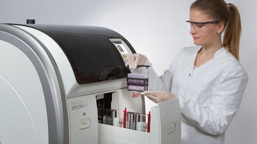
Overview

Product SKU: Workstation Kit - 14051254355, ST - 14051254354, CV - 14051454200
Faster Workflows and Proven Slide Staining Consistency from Slides 1 to 1,600

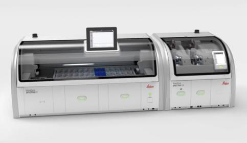
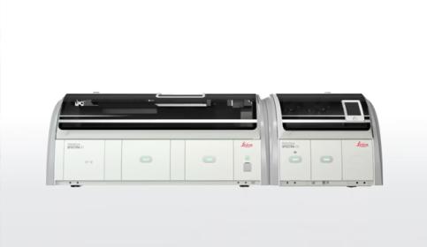
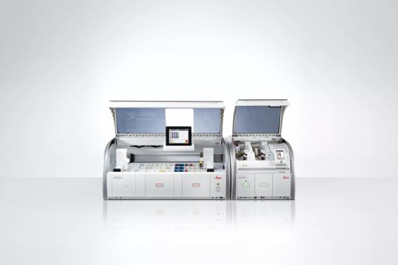
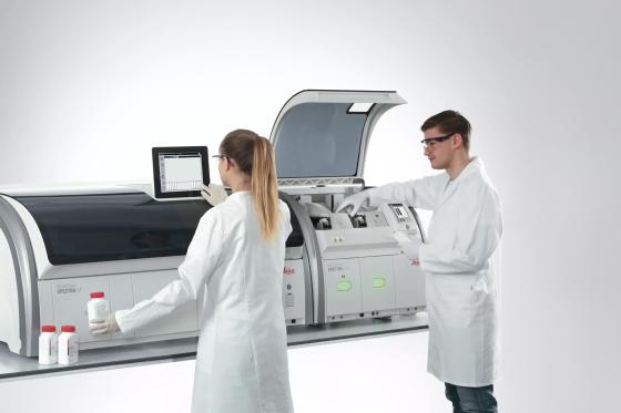
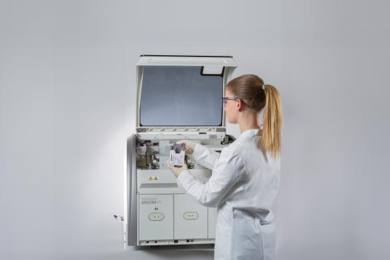
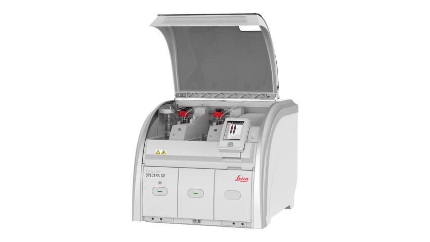
Your time matters. Speed up slide processing while maintaining quality and consistency with the HistoCore SPECTRA Workstation, the only workstation that provides consistent staining from slides 1 to 1,600. It also offers the fastest glass coverslipping drying time on the market at only 5 minutes, processing slides that are instantly dry to the touch.

  • Work smarter: Process up to 570 dried slides per hour, so you can transport immediately and not delay diagnosis.
  • Improve efficiency by 20%: This solution provides the unique flexibility to process H&E and special stains simultaneously at high throughput in parallel with 52 stations.
  • Save time: By using specifically-designed consumables, users ensure consistent staining quality and reduce the number of manual corrective actions for coverslipped slides.
  • Increase confidence: The HistoCore SPECTRA barcode reader effortlessly automates the slide tracking process, freeing up time and resources for other critical tasks.
Contact Us to get a quote. Call +33 9 75 18 50 99 to speak with a representative.

For in vitro diagnostic use.


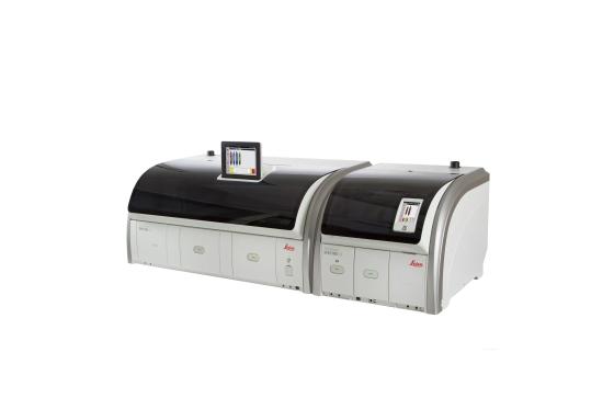
HistoCore CHROMAX WS Workstation for Slide Staining and Coverslipping


HistoCore SPECTRA ST Fully Automated Slide Stainer Features

Automated Routine & Special Staining with Dual Robotic Arms

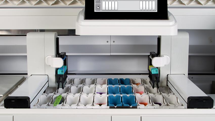
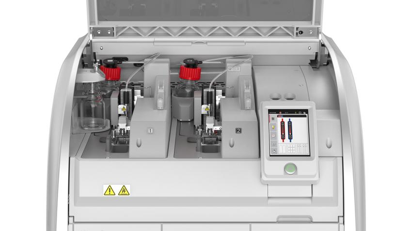
Dual robotic arms and multiple reagent stations ensure each rack is stained efficiently. Users can process up to 360 slides per hour without compromising flexibility or lowering the turnaround time. In addition, the innovative design and 52 cuvette stations enable H&E and special stains to run simultaneously at a high throughput.

Image
Automated Routine & Special Staining with Dual Robotic Arms

Integrated Slide Drying

For better specimen adherence to the glass slide, baking/drying is integrated into the staining process on the HistoCore SPECTRA ST with a built-in hot air oven that eliminates excess paraffin. No external oven or additional user interaction is required, and the oven can process up to six racks at the same time. That means faster turnaround times and quicker processing.

Image
No External Oven Needed

Precise Staining Reagent Management and Cost Control with Unique Single Slide Counting

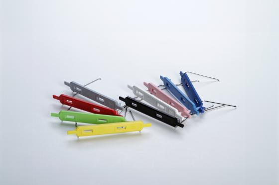
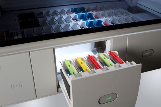
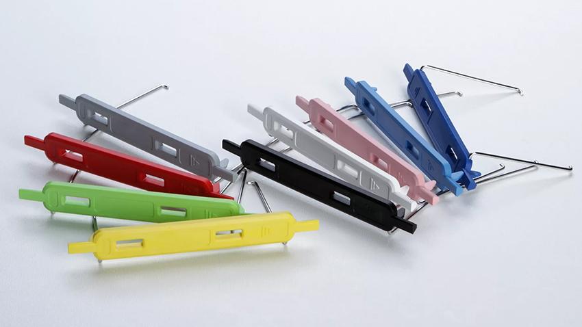
Have precise control over reagent conditions, and coordinate reagent exchange using RFID-monitored consumables, color-coded rack technology, and a unique slide counter. Together, these features minimize user errors, save time, control costs, and reduce waste in the lab.

Image
Precise Reagent Management and Cost Control with Unique Single Slide Counting

Increased Confidence In Your Histology Staining Workflow

Knowing where your slides are improves workflow control and efficiency. The HistoCore SPECTRA barcode reader* effortlessly automates the slide tracking process, freeing up time and resources for other critical tasks. Integrate with your LIS for seamless workflow management. Gather real-time information by automatically scanning slides at the beginning of the staining process, giving you increased confidence in your primary staining workflow.

VIEW 3D ANIMATION

*The HistoCore SPECTRA barcode reader from Leica Biosystems may not be available for use in your country. Please contact your Leica Biosystems representative for more information.

Image
HistoCore_SPECTRA_barcode_reader

Intuitive User Interface Minimizes Hands-On Time

The easy-to-use software of the HistoCore SPECTRA ST automatically calculates optimal bath layouts, eliminating manual steps. As a result, no hands-on interaction is needed, which helps improve lab efficiency.

Image
Intuitive User Interface Minimizes Hands-On Time

Reduced User Errors with Automated Staining Protocol Start

The patented color-coded rack technology provides a clear visual distinction and automatically starts individual protocols when the racks are inserted. This reduces human error by making it easy to recognize and correctly allocate the racks and slides to their designated staining protocols.

Greater Control: Track Staining Reagent Usage, Slide Volume & More

HistoCore SPECTRA H&E Staining System provide the clarity and consistency you expect. In addition, RFID technology enables you to accurately track reagent usage, slide volume, and maintain records.

HistoCore SPECTRA CV Coverslipper Features

Market-Leading Throughput and Drying Time

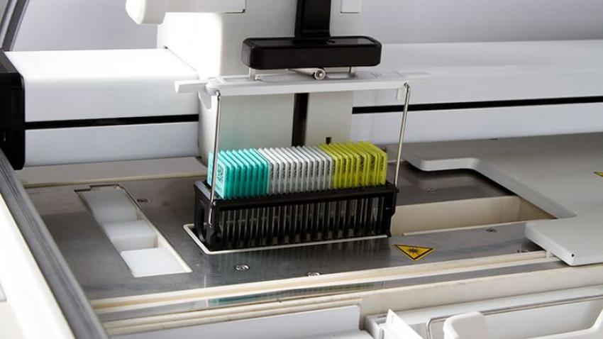
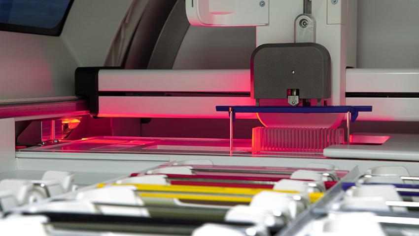
The first and only solution in the market with dual glass coverslip lines enabling the highest throughput: up to 570 dried slides per hour. With an integrated hot air drying oven, it achieves a drying time of only 5 minutes, providing the fastest turnaround time for your lab.

Image
Market-Leading Throughput and Drying Time

Boost Coverslipping Throughput and Efficiency

The dual and independent coverslip lines don’t just deliver the highest throughput, they also give users the flexibility to manually load slides in a continuous workflow. In addition, the coverslipper features an enhanced lay down mechanism, a smooth handling process (push method for coverslipping readiness), and validated ready-to-use consumables with RFID technology. These consumables include the HistoCore SPECTRA CV X1 rapid drying, xylene-based mounting media. An antioxidant is added to inhibit stain fading and prevent the formation of annual rings. It sets up quickly, allowing slide preparations to be handled immediately after mounting.

Image
Boost Coverslipping Throughput and Efficiency

Large Workload Capacity

The 9-rack output drawer can handle a total capacity of 270 slides. That allows users to focus on the many other important tasks throughout the lab.

Image
Large Workload Capacity

Never Worry about Broken Glass

The unique glass detection sensor automatically removes and transfers broken coverslips to better protect the safety of the user while handling slides and to help maintain the integrity of tissue specimens.

Capability to Handle Multiple Labels

The new coverslipping process supports smooth handling of different slide labels, providing flexibility you demand.

Consumables

Achieve Consistent Quality from Slide 1 to 1,600

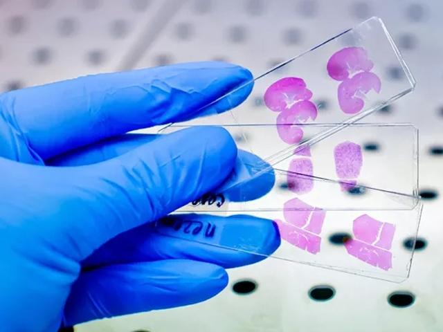
Rest easy knowing the slides processed are of consistently high quality. Leica Biosystems’ validated protocols and the HistoCore SPECTRA H&E Staining System eliminate day-to-day variation. Better yet, these H&E stains provide the clarity and definition to help speed diagnostic efficiency for pathologists and patients.

Image
Achieve Consistent Quality from Slide 1 to 1,600

Ready-to-Use Coverglass and Mounting Media

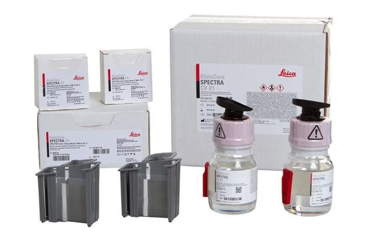
HistoCore SPECTRA CV X1 is a rapid drying, xylene based mounting media. An antioxidant is added to inhibit stain fading and prevent the formation of annual rings. It sets up quickly, allowing slide preparations to be handled immediately after mounting. HistoCore SPECTRA CV X1 is soluble in xylene, toluene, and most xylene substitutes.

HistoCore SPECTRA CV Coverglass allows for maximum reduction of fingerprints due to ready-to-use coverglass cartridges. Enhanced packaging reduces broken coverglass and humidity.

Image
Ready-to-Use Coverglass and Mounting Media

Specifications

HistoCore SPECTRA ST Stainer
HistoCore SPECTRA CV Coverslipper
HistoCore SPECTRA H&E Staining Systems
HistoCore SPECTRA CV Consumables

Configurations

Workstation
Stainer
Coverslipper
Product configuration codes
Workstation Solution
Stainer Only
Coverslipper Only
HistoCore SPECTRA ST Stainer (14051254354)
HistoCore SPECTRA CV Coverslipper (14051454200)
HistoCore SPECTRA CV Cover Glass (3800152) and Mounting Media (3801733)
HistoCore SPECTRA Workstation Kit / Transfer Unit (14051254355)

Resources

Customer Testimonials

Image
Histocore_SPECTRA_CV_Coverslipper_Reduces_Drying_Completion_Time

HistoCore SPECTRA CV Coverslipper reduces turnaround time by 47% from glass coverslipping to slide drying process completion.

An independent university pathology hospital compared the HistoCore SPECTRA CV Coverslipper with the Sakura Tissue Tek® Film Coverslipper. Result: The use of the built-in oven reduced the drying process time by 47% compared to using the drying station of their existing film coverslipper.

Read more

Image
Wisconsin_Diagnostic_Laboratories_Improves_Stain_Quality_and_Workflow

"The fact that the HistoCore SPECTRA ST stainer and stains maintains the same quality from the first slide to the sixteen-hundred is amazing to me.”

Wisconsin Diagnostic Laboratories Histology Department in Milwaukee, Wisconsin, handles more than 24,000 cases and 500,000 blocks every year. They needed to update their staining equipment after 20 years of service and looked for a replacement that would continue their long tradition of highly accurate, efficient service.

Read more

Image
Inform_Diagnostics_teams_up_with_Leica_Biosystems

“The nuclear detail is of excellent quality, much like looking at a picture in a book. The pathologists just love the quality of the slides.”

Always looking to improve its quality and streamline operational efforts, Inform Diagnostics teamed up with Leica Biosystems to assess whether the HistoCore SPECTRA systems provided a cost-effective update to Inform Diagnostics’ staining technology that would meet or exceed its industry-leading quality standards.

Read more

Similar Products

Leica Biosystems BOND-III

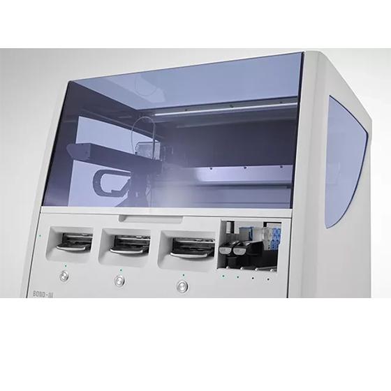
BOND-III Fully Automated IHC and ISH Staining System

Make a Difference with Accurate and Timely Slides

Knowledge Hub

From tissue processing to Digital Pathology, our solutions empower labs with efficiency, precision, and reliability.

the he stain a lesson on consistency and reproducibility
The H&E Stain – A Lesson on Consistency and Reproducibility

Producing an H&E stained slide is a process. It starts way before the slide is loaded on a stainer or moved down through containers by hand. Producing quality, consistent and reproducible H&E stained slides is a process as well as a lesson in...

KP_Hero_Image_TANGERINE_01-640x480
Special Stain Techniques for the Evaluation of Mucins

Special stains that are used for the evaluation of mucins, mucin-like molecules and other carbohydrate containing macromolecules remain in demand and are utilized frequently in the histology laboratory.

H-E-Staining-Overview-A-Guide-to-Best-Practices-640x480
H&E Staining Overview: A Guide to Best Practices

For routine diagnosis, the use of Hematoxylin and Eosin (H&E) is by far preferred for viewing cellular and tissue structure detail by pathologists. 

KP_Hero_Image_MERLOT_01-640x480
An Introduction to Routine and Special Staining

Routine (or H&E) and special stains allow us to visualize otherwise transparent tissue under a microscope and are critical for tissue-based diagnosis.