Breast Macrodissect AI
Developed by Indica Labs, Breast Macrodissect AI is a deep learning tool designed to streamline macrodissection workflows for breast cancer by quantifying tumor content to guide ROI selection and enhance downstream molecular analysis research. Breast Macrodissect AI empowers labs with a high-throughput, user-friendly workflow to maximize efficiency.
Breast Macrodissect AI is For Research Use Only and not intended for clinical diagnostic use. Breast Macrodissect AI is accessed via the Aperio HALO AP image management system.

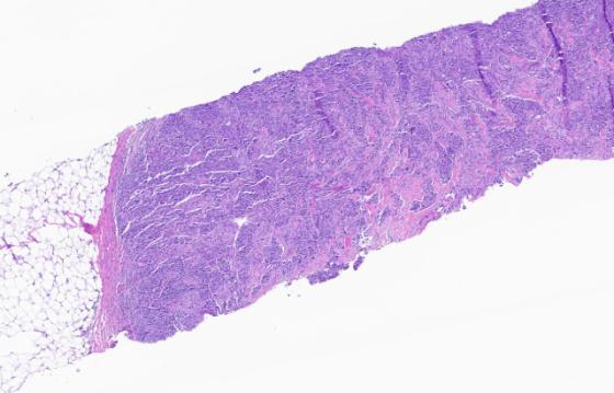
Step 1: Breast Cancer H&E Image
Breast Macrodissect AI adds precision, efficiency, and standardization to the molecular research workflow through the automated tumor content reporting of breast cancer resections, excisions, and biopsy samples.
The algorithm can be launched by a user directly in the viewer or configured to automatically run for every breast H&E image in Aperio HALO AP IMS.
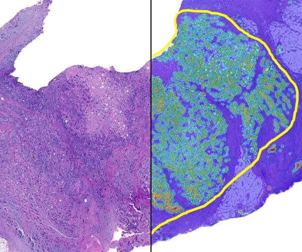
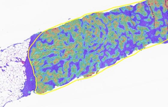
Step 2: Tumor Density Heatmap
A cancer cell density heatmap is displayed allowing identification of the optimal region for downstream macrodissection. The yellow boundary identifies this area.
Prior to this step, image analysis pre-processing excludes artifact and benign epithelial tissue and segments regions of necrosis.
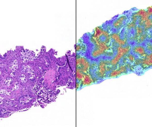
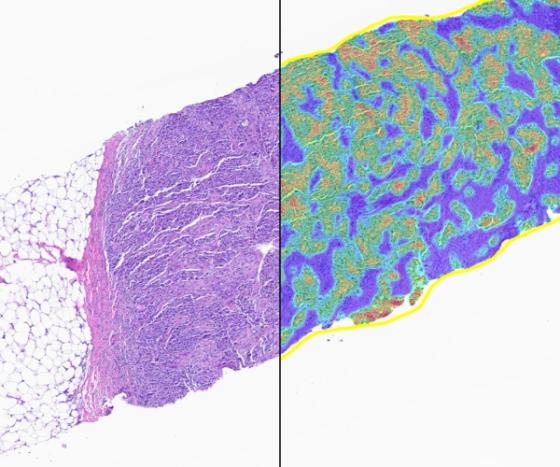
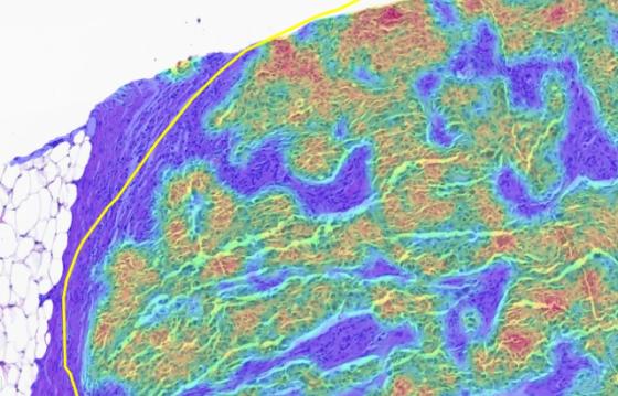
Step 2: Tumor Density Heatmap (Higher Resolution)
A cancer cell density heatmap is displayed allowing identification of the optimal region for downstream macrodissection. The yellow boundary identifies this area.
The warmer the heatmap color, the higher the density of cancer cells.
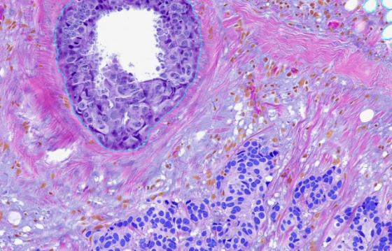
Step 3: Cancer Cell Classification
Each individual cell across the whole slide image is classified as cancer or other. Precise and standardized tumor content is reported across the whole sample and within the region selected for macrodissection.
- Cancer Cells: Blue
- Other Cells: Orange
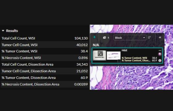
Step 4: Breast Macrodissect AI results
Whole slide and dissection area tumor content are displayed in the slide tray and assay window.
Breast Macrodissect AI outputs the following results:
- Whole Slide Image
- Total Cell Count
- Tumor Cell Count
- % Tumor Content
- % Necrosis Content
- Dissection Area
- Total Cell Count
- Tumor Cell Count
- % Tumor Content
- % Necrosis Content
Specifications
| File Formats: | Non-proprietary (JPG, TIF, OME.TIFF, DICOM [DCM]), Leica (SVS, AFI, SCN, LIF), Hamamatsu (NDPI, NDPIS), Philips (iSyntax, i2Syntax), 3DHistech (MRXS), Nikon (ND2), Akoya (QPTIFF, component TIFF), Olympus / Evident (VSI), Zeiss (CZI), Ventana (BIF), KFBIO (KFB, KFBF) |
| IMS: | Aperio HALO AP IMS |
| Algorithm: | Breast Macrodissect AI |
| Tissue Type: | FFPE histology whole tissue resections and biopsy of primary and metastatic breast cancer (adenocarcinomas) |
| Scoring: | Tumor Cell Percentage |
| Regulatory status: | Research Use Only (RUO) |
Breast Macrodissect in Aperio HALO AP IMS Demonstration
Explore the future with the Breast Macrodissect AI app from Indica Labs. Breast Macrodissect AI seamlessly integrates within the Aperio HALO AP IMS enterprise digital pathology platform to enhance the molecular research workflow through automated tumor content reporting of primary invasive breast carcinoma resections, excisions, and/or core needle biopsies.
After selecting an image, view the H&E slide in the viewer and the Breast Macrodissect AI results. Results include overlays and quantitative results. The first set of overlays prepare the tissue for analysis by identifying areas of analyzable tissue. The next overlay identifies areas of tumor in the tissue, with the tumor density heatmap highlighting the densest areas of tumor. With the assistance of the heatmap, users identify the dissection area by drawing a region of interest with immediate quantitative feedback on the number of all cells and tumor cells in the region to ensure proper sampling.
Related Product Applications